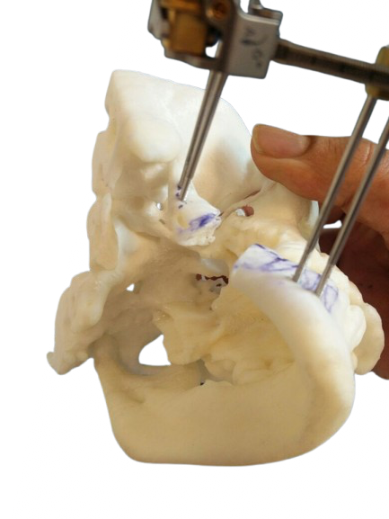
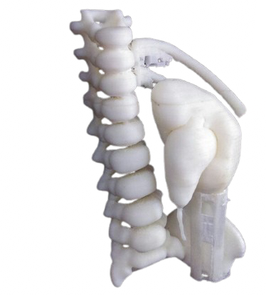
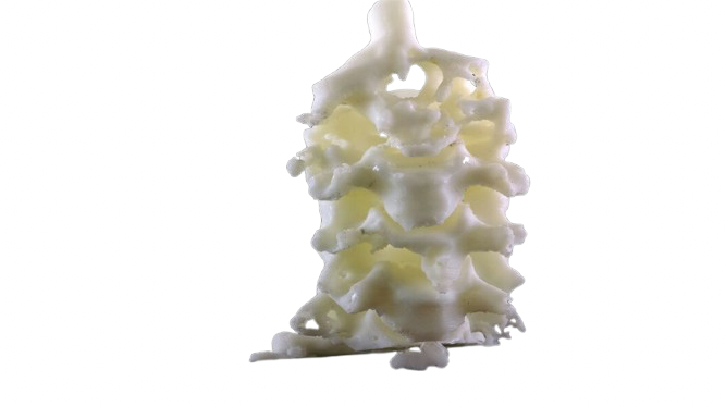
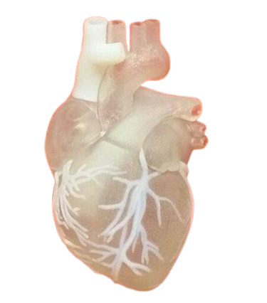
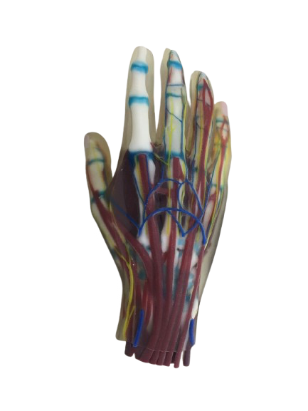
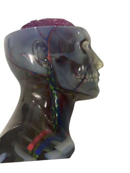
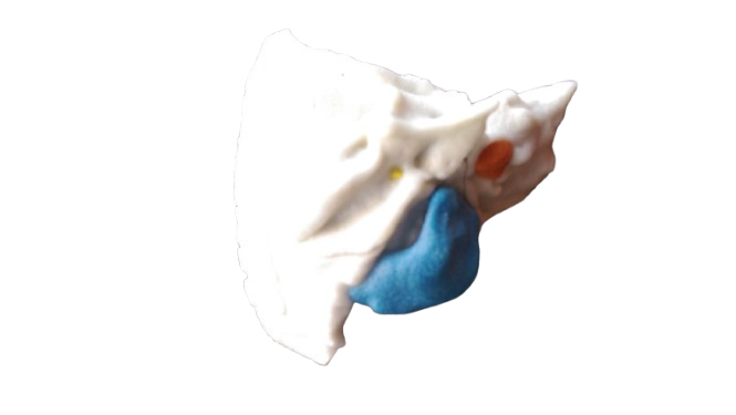
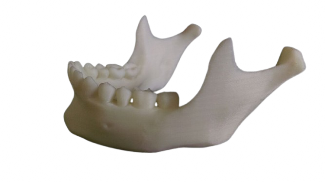
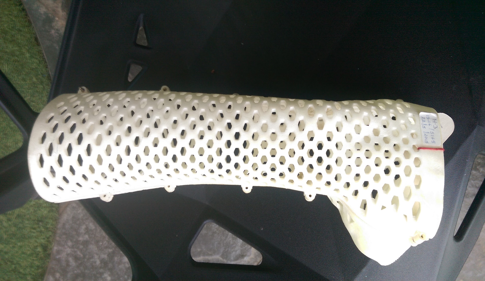
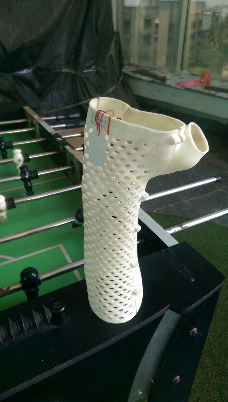
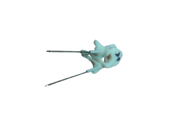
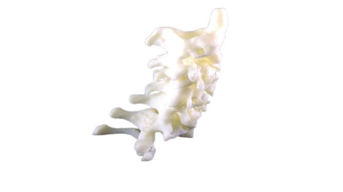
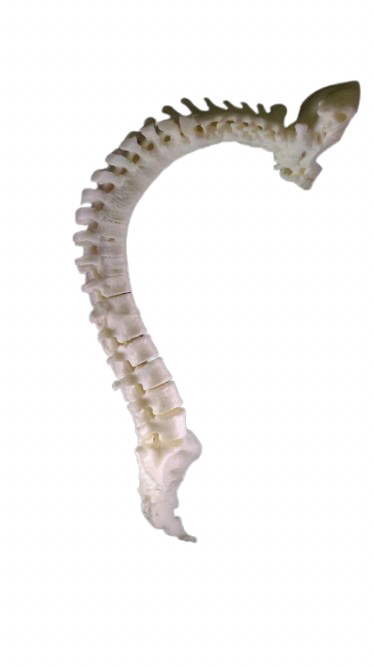
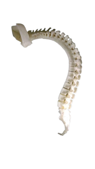
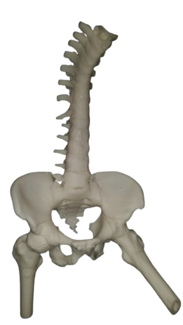
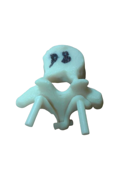
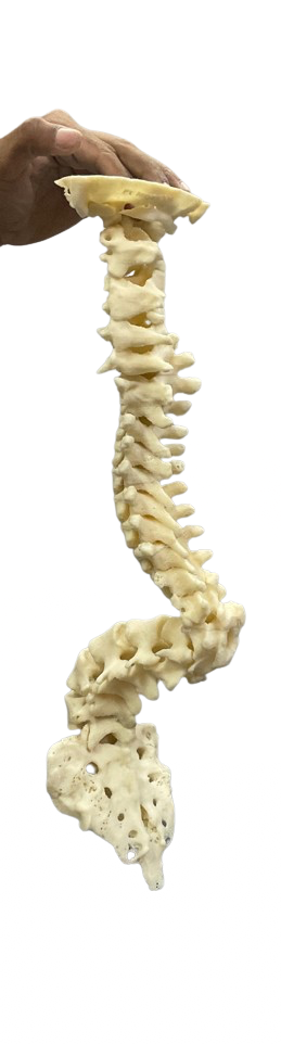
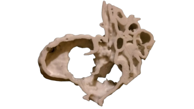
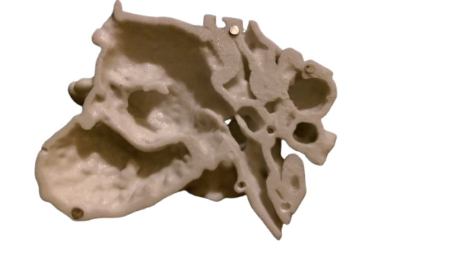
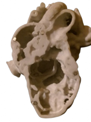
3D printed anatomical models provide high-fidelity, patient-specific replicas derived from DICOM imaging through advanced DICOM-to-3D workflows. These surgical planning models enable detailed pre-surgical visualization, offering critical insights for complex craniofacial, orthopedic, and oncological procedures. By accurately replicating anatomical structures, the models support case simulation, intraoperative referencing, and interdisciplinary planning. They serve as valuable educational 3D anatomy tools, enhancing surgical training and resident instruction through tangible, real-scale models. In clinical practice, these patient-specific medical models facilitate clearer communication among surgical teams and improve patient comprehension of planned interventions. Their use contributes to reduced intraoperative uncertainty and greater procedural confidence, particularly in challenging reconstructions and tumor resections. As custom surgical training tools, they bridge the gap between imaging and operative execution, reinforcing precision in both planning and performance.